9. Advanced/Optical sectioning illumination modalities#
9.1. Confocal microscopy#
9.2. TIRF#
9.3. Light sheet microscopy#
Note
If you want to read up more about light sheet microscopy, I recommend the following review:
Stelzer, E.H.K., Strobl, F., Chang, BJ. et al. Light sheet fluorescence microscopy. Nat Rev Methods Primers 1, 73 (2021).
This subchapter on light sheet loosely follows M. Fazel, K. S. Grussmayer, B. Ferdman, A. Radenovic, Y. Shechtman, J. Enderlein, S. Pressé, Fluorescence Microscopy: a statistics-optics perspective, [OA, arXiv:2304.01456].
There are many different optical realizations of light sheet microscopy (LSM) - all of them generate a thin sheet of light to excite the sample only within the focal volume. This is in contrast to previously discussed 3D microscopy modalities like confocal or optical sectioning SIM; they provide sectioning only in detection and still illuminate all the specimen along the optical path of the excitation. TIRF microscopy avoids this unnecessary light dose, but restricts the illumination and thus observation volume to close to the coverslip.
LSMs are ideal to observe dynamic processes in 3D over long periods of time, because photodamage and photobleaching are greatly reduced. They are also a method of choice to image large, optically cleared specimen because they reduce out-of-focus light due to easy optical sectioning. The setups are usually constructed to permit manipulation of the sample in three dimensions and allow observation of living 3D specimens without having to fix them on a flat surface.
The classical modern light sheet implementations illuminate the sample from the side, with the excitation generated by an objective or lens and a Gaussian beam orthogonal to the objective used for detection. The detection path uses a widefield geometry with a camera, and the excitation and detection paths are uncoupled.
Stelzer et al. coined the term selective-plane illumination microscopy (SPIM), which is often used as synonym for LSM. They used a cylindrical lens to form an astigmatic Gaussian beam, effectively elongating the beam along one dimesion to generate a thin, static light sheet. Soon after, digitally scanned laser light sheet microscopy (DSLM) came into play. Here, the LS illumination is achieved by rapidly scanning in one direction during the camera exposure/synchronized with the rolling shutter; this is usually accomplished with galvanometric mirrors.
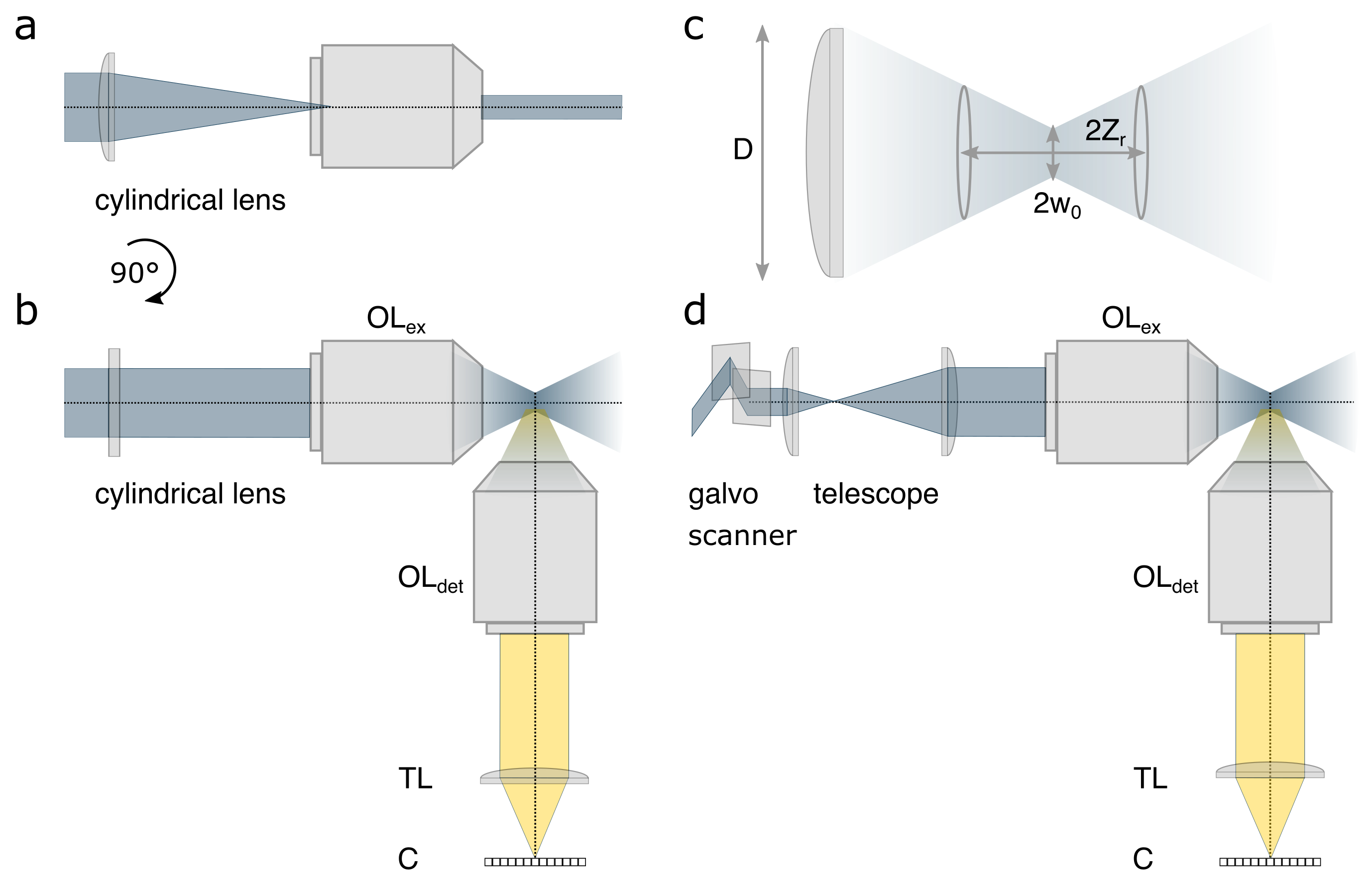
Fig. 9.1 a) In SPIM, a static light-sheet is formed by a cylindrical lens in the excitation path creating an elongated beam in one direction focused in the sample through the excitation objective lens (\(\text{OL}_{\text{ex}}\)). b) the same perpendicular detection optics as in panel a). c) A schematic of the Gaussian beam in panels a-b focused through a lens or objective with diameter D, beam waist \(\omega_0\) and Raleigh length \(z_r\). d) In Digitally scanned laser Light-Sheet Microscopy (DLSM) a galvanometric (galvo) scanning unit rapidly moves a Gaussian beam perpendicular to the detection axis focused in the sample through the excitation objective lens (\(\text{OL}_{\text{ex}}\)). Signal from the focal plane is collected through the detection objective lens (\(\text{OL}_{\text{det}}\)) and tube lens (TL) onto a camera ©.#
For the most simple estimation of the axial resolution of LSM, the extent of the light sheet or the thickness of the Gaussian beam at twice the beam waist \(R_{axial} = 2 w_0\) can be used. Similarly, twice the Raleigh length \(2z_r\) provides a measure of the field-of-view.
where \(w_0\) is the beam waist, \(\theta\) is the, f and D are the focal length and the diameter of the lens, NA is the numerical aperture and \(n\) the refractive index of the medium and \(\lambda\) the wavelength of the excitation laser.
Thinner light sheets lead to a better axial resolution, but they achieve worse uniformity of illumination across FOV and vice versa. Most commonly, one photon fluorescence illumination is used, which is ideal for developmental biology due to low phototoxicity, but also 2-photon, Raman spectroscopy or others can be applied as imaging contrast mechanisms.